Use of Gold probe
The module covers:
- When to use Gold probe
- Different parts of the Gold probe
- How it is set up and used
- Other types of thermal devices in use
When to use Gold probe
- Mainly in peptic ulcer bleeds
- Bleeding polyp stalks
- Dieulafoy lesions
- Mallory-Weiss tears
- Arterioveous malformations (AVMs)
Different parts of the Gold probe
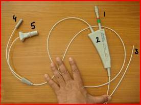
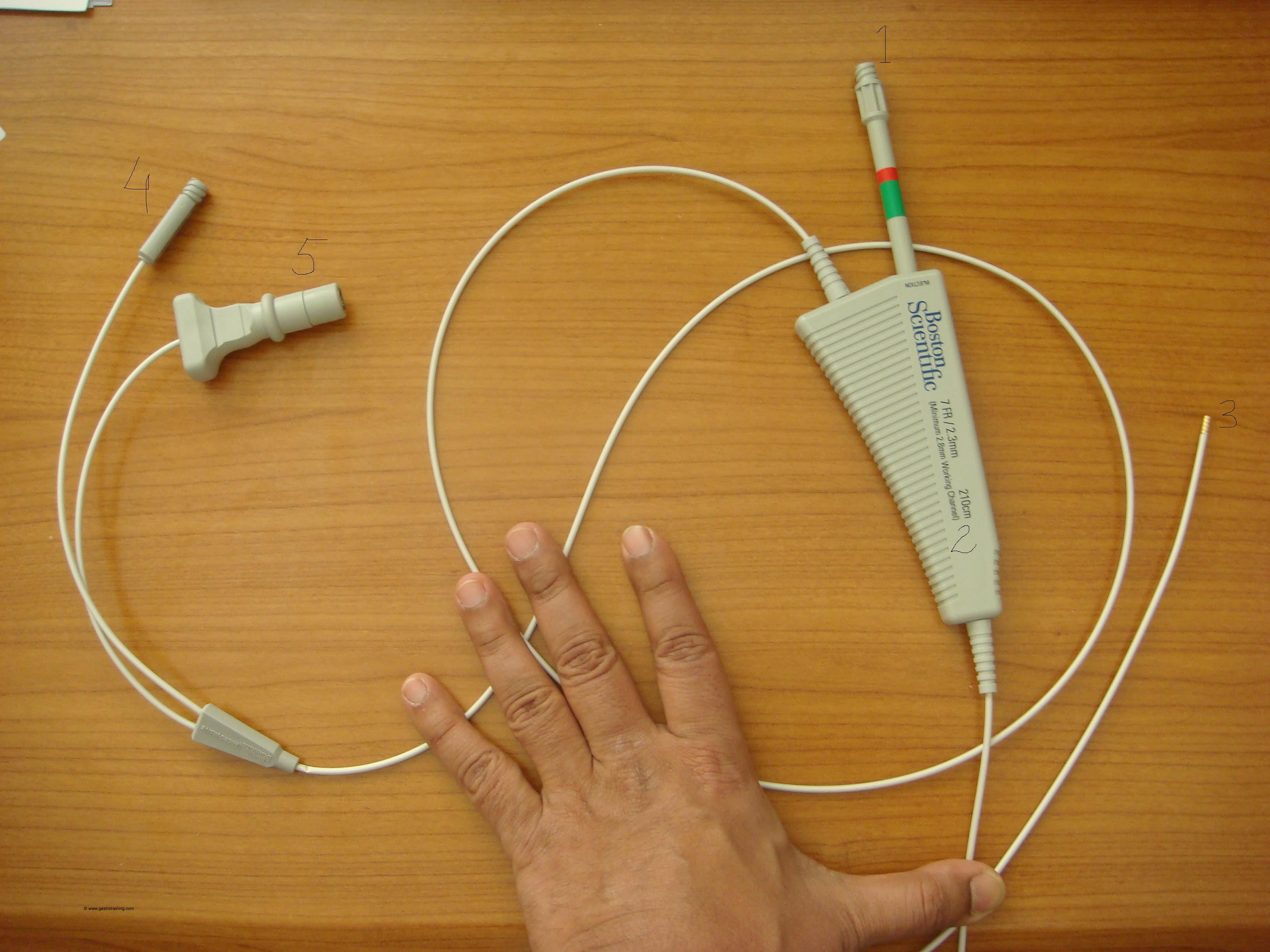
Injection Gold Probe Catheter can be used to give injection therapy and also for electro haemostasis. It has also got irrigation capabilities.
Picture1: The gold probe
|
- The catheter handle is a thick triangular portion- from its apex emerges the cable leading to the gold tip
- From the base of the catheter handle arises
- The injection hub (with the red/green mark) and
- The cable which splits into two further cables
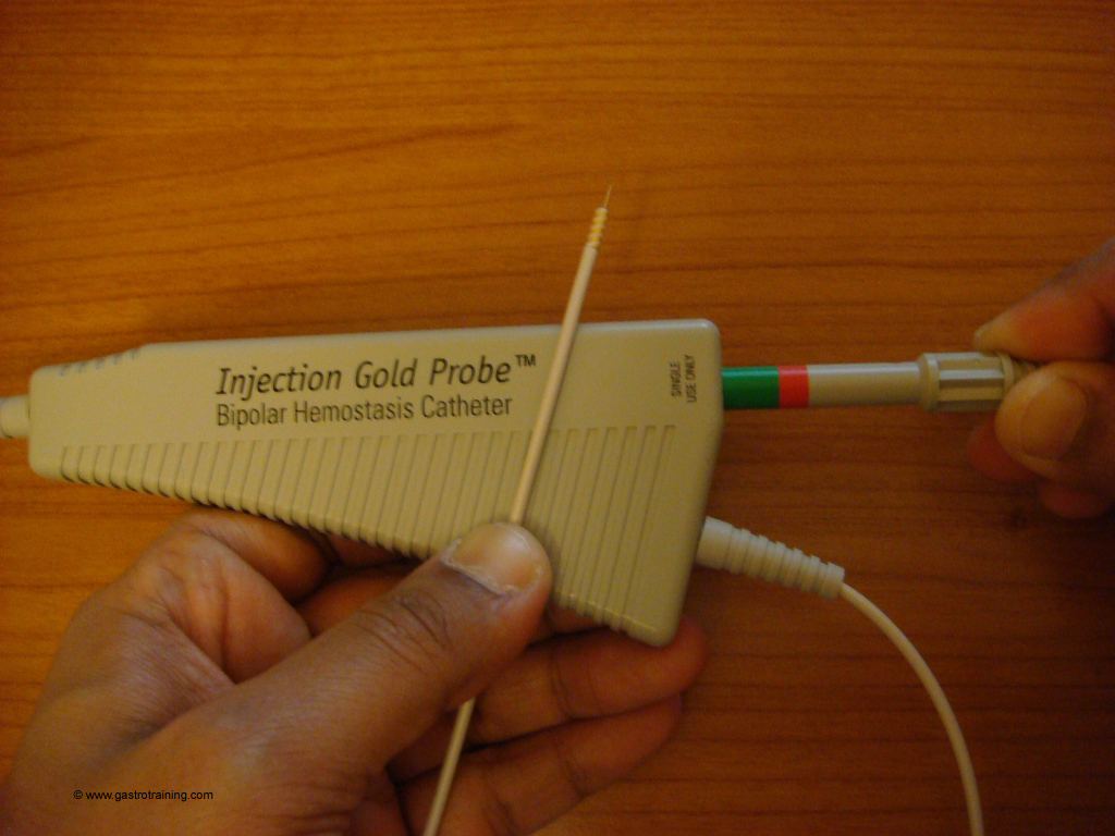
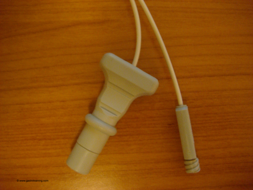
- One with the thicker, flange shaped end is the bipolar electrical connector- which is connected to the cable coming from the bipolar socket of the ERBE diathermy box (ICC 200)
- The other cable takes a saline filled syringe to flush the tip after burning but alternatively you can exchange the adrenaline syringe with a saline filled syringe and flush.
Picture2: The electrical connector/ irrigation hub
- The device is supplied in 7Fr (2.3mm) and 10Fr (3.2mm)- 7F and 10F probes require a minimum of 2.8mm and 3.7mm working channels respectively. So use 7F Gold probe if your endoscope is not a therapeutic one (yellow colour as opposed to salmon colour which is therapeutic)
- The length of the gold probe is usually 210cm but 300cm and 350cm is also available to use in particularly deep in small intestine and colon if needed.
How is it set up and used
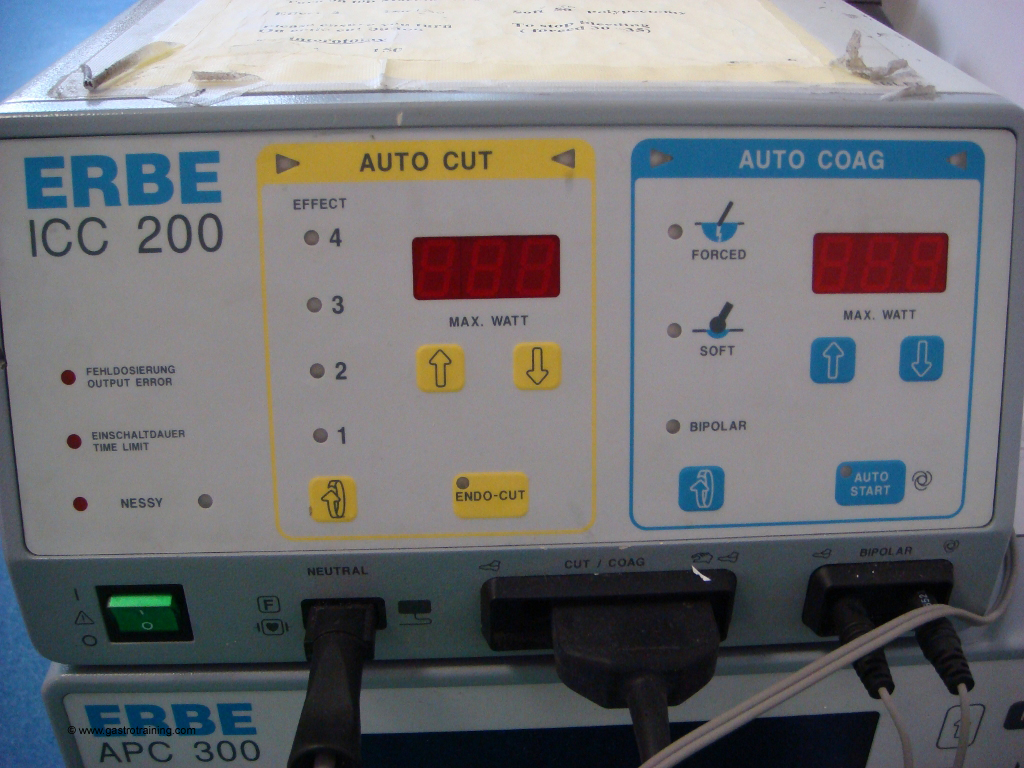
- Connect the bipolar electrode end to the bipolar socket of the ERBE box
- For gold probe:
- No patient plate/neutral cable is needed ( in the picture it is left connected from previous use but is not used)
- Accessory cable (gold probe in this case) is attached to the bipolar socket in the diathermy box (ICC 200)
- Nothing goes in the cut/coag socket (the middle socket in the diathermy box- we just left the plug of the APC in from previous use but it is not needed)
- Cutting panel is not needed at all and the setting on the autocut panel is irrelevant and the yellow pedal should not be used.
- Chose Autocoag with bipolar effect (as opposed to soft or forced which we used before), power to 15-30W for visible vessels/ Dieulafoy lesion/ Mallory Weiss lesion
- Choose power to 10-15W for colonic bleed (AVM/ diverticular bleed)
- Connect a saline filled syringe to the irrigation hub and inject water until water is visible at the distal tip of the probe
- Test the probe before passing it through the endoscope by touching the tip to a 1-2ml of saline / KY jelly and depressing the footswitch to activate the probe tip- saline bubbles are to be seen and steam should be emitted
- Adrenaline filled syringe (1:10000 dilution adrenaline) is attached to the injection hub and pull back on the injection hub until hub locks into position to ensure that the injection needle is completely retracted into the probe tip
- Turn off the electrosurgical generator during the insertion of the Gold Probe
- Advance the tip until the gold tip is endoscopically visible through the endoscope
- For lesions in the duodenum sometime you might find resistance in passing the gold probe when it’s tip reaches tip of the scope- then straighten the scope as much as possible and try again.
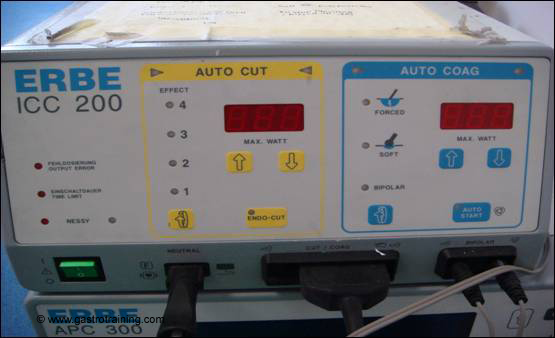
Picture3: The ERBE diathermy box ( ICC 200)
To use the gold probe to inject adrenalin
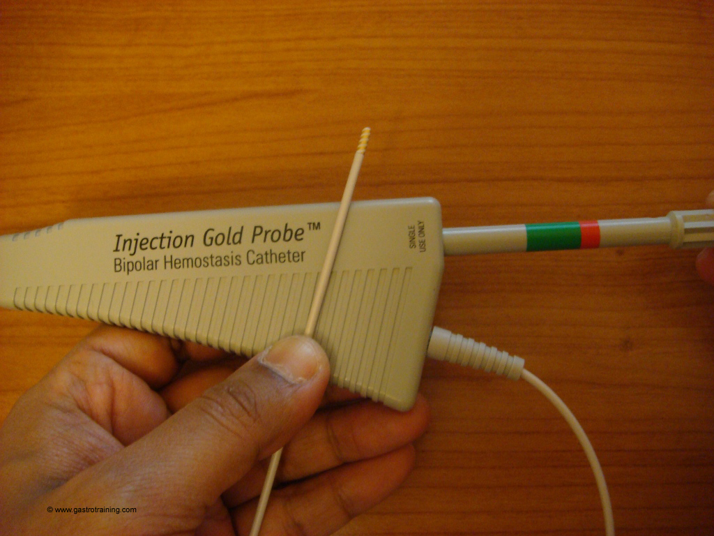
- After positioning the tip near the lesion – slowly push the injection hub to the catheter handle until full extension of the needle is visible (4-6mm)
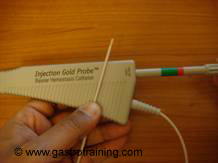
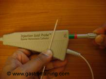
Picture4: Pushing the handle in makes the needle to come out of the sheath - Under typical endoscopic configurations the green band on the injection hub should be partially visible.
- Under some very tortuous configurations the green band and/or the red band may be completely hidden. But never push the injection hub past the proximal end of the red band
- Insert the extended needle into the selected site and inject 1:10000 dilution adrenaline in 2-3ml aliquots and then completely withdraw the needle once finished.
- Remember the volume of the adrenaline solution is important to exert tamponade effect.
To use the gold probe for electrohaemostasis
- Identify and position the endoscope proximal to the intended cautery site.
- Advance the probe until perpendicular or tangential contact is made with the site. Good apposition of the tip to the tissue is important (co-aptive pressure)
- Using the Blue foot pedal activate the tip to cauterize the site- 2-5secs
- Irrigate with saline before detaching the tip from the burnt area to avoid sloughing of devitalized tissue.
Other types of thermal devices in use
A) Heater probe ( Unipolar) – Teflon coated hollow aluminium cylinder with inner heating coil- heats tissue directly
B) Bipolar (Multipolar) – generates heat indirectly by passage of electric current. Two electrodes in the tip complete a circuit through non-desiccated tissue.
Types
- HEMArrest- Bard interventional products
- Gold Probe- Microvasive, Boston Scientific
- BICAP- Circon Acmi
- Quick silver- Wilson-Cook Medical Inc.
Here is the link for Gold probe Video:
Acknowledgement/Bibliography:
- Gustavo A et al. Thermal probes alone or with epinephrine for the endoscopic haemostasis of ulcer haemorrhage Best Practice & Research Clinical Gastroenterology June 2000 :14 (3): 443-458
- Jensen DM et al.CURE multicenter randomized, prospective trial of gold probe vs. Injection & gold probe for hemostasis of bleeding peptic ulcers. Gastrointestinal Endoscopy 1997:Volume 45 (4), Page AB92
- Arasaradnam RP et al.Acute endoscopic intervention in non-variceal upper gastrointestinal bleeding Postgrad Med J. 2005;81:92-98
- Product guide of the respective companies- Boston Scientific


