Portal vein
Discuss the portal vein formation?
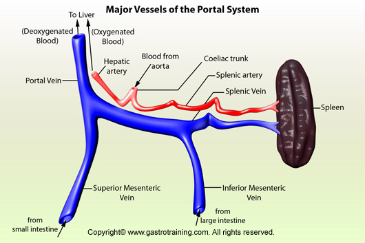
Venous blood from the stomach and small intestine is collected by the portal vein. The portal vein is formed posterior to the neck of the pancreas, by the union of the superior mesenteric and splenic veins. The inferior mesenteric vein ends by joining the splenic vein. The left and right gastric veins drain directly into the portal vein. The right gastroepiploic veins drain into the superior mesenteric vein or splenic vein or directly into the portal vein. The left gastroepiploic vein and short gastric veins drain into the splenic vein.
Discuss portal-systemic anastomoses?
The portal and systemic venous system communicates in several locations. These anastomoses are important clinically. When portal system is blocked, the blood from the GIT can still reach the right atrium through these anastomoses (they dilate and become varicose when portal system is obstructed as well as causing reversed flow of portal blood into systemic veins). The important anastomoses are as follows:
- Gastro oesophageal region-left gastric vein branches anastomose with the oesophageal veins that drain into the azygous vein.
- Anorectal region- superior rectal vein anastomoses with the middle (branch of internal iliac) and inferior rectal vein (branch of internal pudendal)
- Paraumbilical region- Para-umbilical veins in the falciform ligament connect the portal vein with subcutaneous vessels around the umbilicus, and these last drains into the epigastric veins and thence into the venae cavae
- Retroperitoneal region- splenic and pancreatic vein tributaries anastomose with the left renal vein. Numerous, small retroperitoneal veins drain the “bare” (i.e., non-peritonealised) surfaces of the organs also communicate with the veins of the diaphragm.


