Gallbladder (GB)
Discuss the anatomy of GB?
It is a pear shaped organ that lies on the visceral surface of the liver. The GB is divided into a fundus, body and neck. The fundus is the wide end of GB that projects from the inferior border of the liver. The body of the GB lies in contact with the visceral surface of the liver. The neck of the GB is narrow and may have a dilatation called the Hartmann’s pouch. The duct of the GB (cystic duct) joins the common hepatic duct to form common bile duct.
GB is supplied by the cystic artery branch of right hepatic artery
Lymphatic drainage is to cystic node (and then to hepatic and coeliac node)
The GB stores (30-60mls) and concentrates the bile secreted by the liver. The GB contracts and release bile in response to a meal.
Discuss the effects of cholecystectomy?
GB is a vestigial organ like the appendix
Liver normally produces 1-1.5 litres of bile a day. It is constantly produced and thus there is a steady amount of bile entering the duodenum. Some of it enters the GB as it comes down the duct and stored there.
The bile that constantly flows into the duodenum is adequate for fat digestion. Thus cholecystectomy does not lead to fat maldigestion. However, a rich fatty meal can sometimes cause a degree of diarrhoea.
Discuss the entero hepatic circulation for bile acids?
Liver produces bile acids (cholic acid and chenodeoxycholic acid) from cholesterol. These bile acids are conjugated with glycine and taurine to form water soluble primary conjugated bile acids (also called bile salts). These bile salts are secreted with bile in the intestine. Bile salts are ionised (due to pH of the intestine) and dehydroxylated (by the intestinal bacteria) to from primary and secondary conjugated bile salts (which are still water soluble). These conjugated bile salts are actively reabsorbed in the ileum. Liver extracts bile acids from portal blood and secretes it again in the canaliculi. This is the enterohepatic circulation.
95% of the bile acids which are delivered to the duodenum will be recycled by the enterohepatic circulation. The rest is lost in faeces and replaced by de novo synthesis.
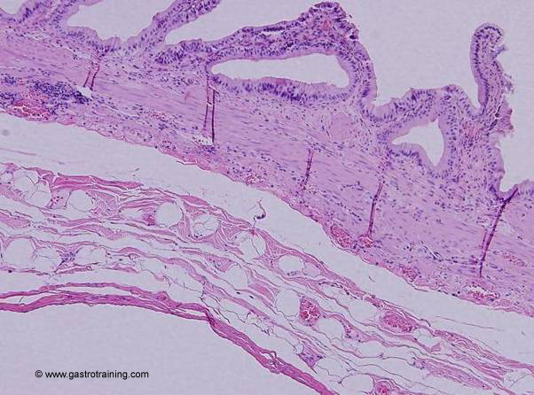
Pic 1: Gallbladder wall: showing all layers from mucosa to serosa. Two Rokitansky-Aschoff sinuses underlie the mucosa in centre & right photo.


